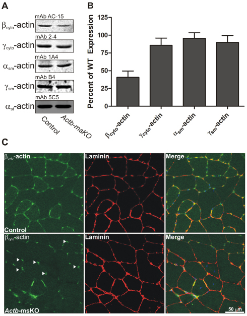
Fig. 1.
Skeletal muscle-specific ablation of βcyto-actin. (A) Representative western blots of actin isoform expression from actin-rich eluates of skeletal muscle from control and Actb-msKO mice. (B) Quantification of actin isoform expression from quadriceps extracts from three control (WT) and three Actb-msKO mice at 3 months of age. αst-actin served as a loading control. Actb-msKO skeletal muscle showed a 59% decrease in βcyto-actin expression. Error bars represent s.e.m. (C) Cryosections of 10 μm from control and Actb-msKO quadriceps were stained with DAPI (blue), βcyto-actin (green) and laminin (red). Control skeletal muscle showed sarcolemmal staining, which was absent in Actb-msKO skeletal muscle. Endomyosial capillaries (arrowheads) showed strong βcyto-actin immunoreactivity, probably explaining the remaining βcyto-actin signal in actin-rich elutes of Actb-msKO muscle. Scale bar: 50 μm.

