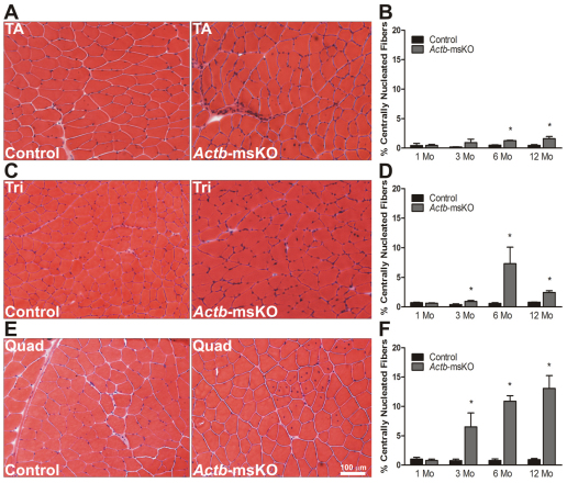
Fig. 2.
Quadriceps myopathy in Actb-msKO mice. (A–F) Representative 10 μm sections stained with hematoxylin and eosin, and respective quantification of the proportion of centrally nucleated fibers from control and Actb-msKO tibialis anterior (A,B), triceps (C,D) and quadriceps (E,F) muscles at 1, 3, 6 and 12 months of age (n=3 mice per genotype per timepoint). *P≤0.05. Error bars represent s.e.m. Scale bar: 100 μm.

