
Fig. 1. Identification of CMV-reactive CD8+ T cells in peripheral blood lymphocytes from M. nemestrina.
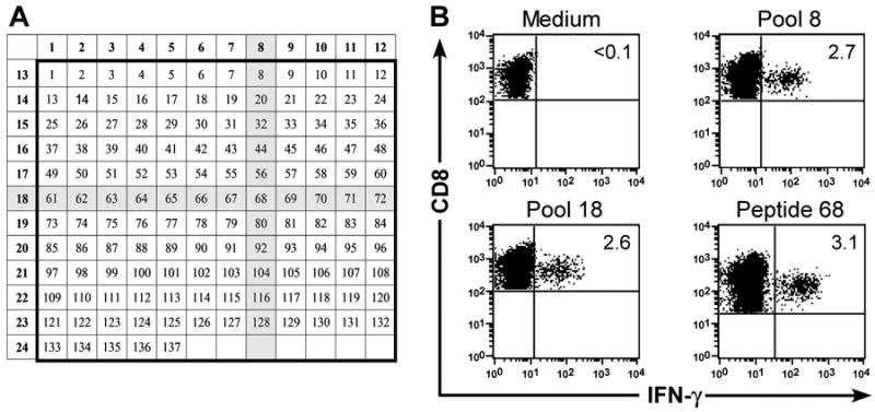
(A) Arrangement of 137 15-mer peptides spanning the sequence of the rhCMV IE-1 protein. The shaded areas correspond to the peptides present in the two positive pools (8; 18) in a representative epitope mapping experiment. PBMC obtained from a 23 macaques were examined for the presence of CMV-specific T-cell responses by CFC. (B) CFC detects IFN-γ production by CMV-specific CD8+ T cells after stimulation of PBMC with CMV IE-1 peptides in pool 8 (upper right panel) and 18 (lower left panel). Stimulation of PBMC with the corresponding peptide 68 (lower right panel) confirmed that sequences within the single peptide shared by both pools 8 and 18 stimulated IFN-γ-production by CD8+ T cells. PBMC stimulated with medium alone (upper left panel) served as a negative control. Data are gated on CD3+CD8+ cells.