
Fig. 2. Derivation of macaque CMV-specific CD8+ T cells from TEM or TCM subsets.
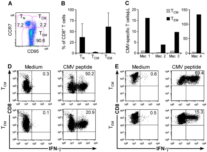
(A) Flow cytometry showing CD8+ T-cell subsets in macaque PBMC including TCM (CCR7+CD95+), TEM (CCR7−CD95+), and TN (CCR7+CD95−). (B) Frequency of CD8+ TCM, CD8+ TEM, and CD8+ TN (%) in peripheral blood CD8+ lymphocytes. Aliquots of PBMC obtained from 18 healthy macaques were stained with mAbs binding to CD8, CD3, CD95, and CCR7 and examined by flow cytometry after gating on CD3+CD8+ T cells. Mean and SD is shown. (C) CMV IE-specific T cells are present in distinct CD8+ TM subsets. CFC assay for IFN-γ+CD8+ T cells specific for CMV in sort-purified CD8+CD62L+ and CD8+CD62L− T-cell subsets obtained from 4 macaques. The absolute number of CMV-specific CD8+ TCM or TEM/μL peripheral blood was determined by calculating the absolute number of CD3+CD8+ T cells/μL of blood (% of CD8+ T cells/lymphocyte subset × lymphocyte count/μL blood/100). Subsequently, the absolute number of the TEM, TCM, and TN subset was derived (% subset × number of CD3+CD8+/μL blood/100). Finally, we calculated the absolute number of CMV-specific CD8+ T cells/μL in each subset (% IFN-γ+ cells × absolute number of CD3+CD8+ TCM+TN/μL or CD3+CD8+ TEM/μL blood/100). (D, E) CFC assay of macaque CMV-specific T-cell lines detects IFN-γ+CD8+ CMV-specific T cells in sort-purified CD8+CD62L+ cells containing TCM (upper panels) and CD8+CD62L− TEM subsets (lower panels). Sort-purified subsets from 2 representative macaques were stimulated with autologous CMV peptide-pulsed antigen-presenting cells and assayed by CFC for production of IFN-γ after stimulation with medium alone (left panels), or CMV peptide antigen (right panels). Data are gated on CD3+CD8+ cells.