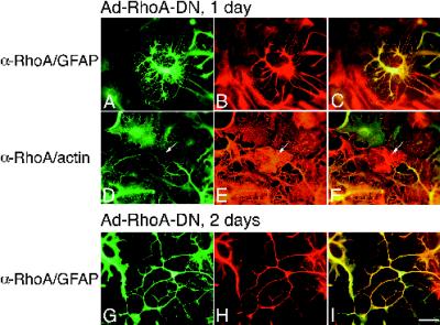
Figure 6.
RhoA-DN induces morphological differentiation of astrocytes. (A–F) Images of cells infected with Ad-RhoA-DN at high m.o.i. for 1 d. (A–C) Images of cells stained with α-RhoA antibody/FL conjugate to recognize infected cells (A) together with α-GFAP/TR conjugate to identify the cells as astrocytes (B). (D–F) Images of cells stained with α-RhoA antibody/FL conjugate (D) together with rhodamine–phalloidin to recognize actin (E). C and F are double exposures. Note that the uninfected cell in the center of the image in D–F (arrow) has actin fibers (E). (G–I) Images of cells infected for 2 d with Ad-RhoA-DN and stained with α-RhoA/FL (G) together with α-GFAP/TR (H). Bar, 25 μm.

