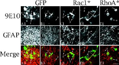
Figure 9.
Effects of Rac1* and RhoA* on process growth in neurons and astrocytes in cultured organotypic hippocampal slices. (A1–A3 and B1–B3) Confocal images of cells from organotypic slices infected for 2 d with Ad-GFP and stained with 9E10/FITC (A1 and B1) together with GFAP/Cy5 (A2 and B2) to identify the cells as astrocytes. Cells labeled “a” (astrocytes) are identifiable in the GFAP/Cy5 channel, whereas cells labeled “n” (neurons) were not. Other experiments confirmed that GFAP-negative cells stained with neuronal markers such as MAP2. Images in A1 and B1 result from GFP fluorescence and not 9E10 staining because uninfected cultures had no signal in any plane in the FITC channel. Images from the top row (A1–E1) were pseudocolored green, and images from the middle row (A2–E2) were pseudocolored red. The pseudocolored images were combined to generate the merged image in the third row (A3–E3). (C1–C3 and D1–D3) Confocal images of cells from organotypic cultures infected for 2 d with Ad-Rac1* and stained as above. Rac1* causes loss of processes on astrocytes (a in C) but not on neurons (n in D). The neurons are not evident in the GFAP/Cy5 channel (D2). (E1–E3) Confocal images of cells from organotypic cultures infected with Ad-RhoA* and stained as above. Note that RhoA* causes loss of processes on astrocytes (a). Insets illustrate the effects of RhoA* on neurons. n in the inset identifies a cell lacking processes that is not evident in the GFAP channel (E2 inset).

