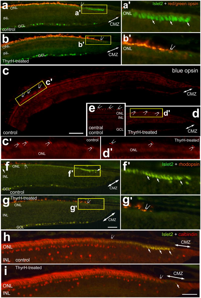
Figure 2.
TH stimulates the expression of opsins and calbindin in photoreceptors in far peripheral regions of the postnatal chick retina. Treated eyes received injections of 2 μg of TH at P0, P1, P2 and P4, whereas control eyes received injections of vehicle. Sections of the peripheral retina and CMZ were labeled with antibodies to Islet2 (green; a, b and f–i), red/green opsin (red; a and b), violet opsin (red; c–e), rhodopsin (red; f and g), and calbindin (h and i). Panel e is a representative image of immunolabeling for violet opsin in the outer segments of cone photoreceptors in a central region of retina, approximately 3 mm from the CMZ. Double-ended arrows indicate the domain of the CMZ, arrows Islet2-positive cells in the ONL, and small double-arrows indicate the onset of expression photoreceptor markers. Areas indicated by yellow boxes are enlarged 3-fold in the panels immediately to the right (a′, b′, f′ and g′). Panels c′ (approximately 1000 μm) and d′ (approximately 300 μm) are 4-fold enlargements of the areas indicated by yellow boxes in c and d. The calibration bar (50 μm) in panel c applies to panels c–e, the bar in g applies to a, b, f and g, and the bar in i applies to h and i. Abbreviations: ONL – outer nuclear layer, INL – inner nuclear layer, IPL – inner plexiform layer, GCL – ganglion cell layer.

