Figure 5.
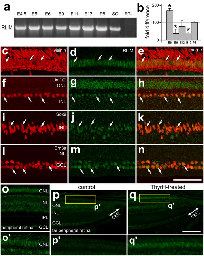
RLIM is dynamically expressed during retinal development, and is up-regulated in mature photoreceptors and in response to treatment with TH. RT-PCR (a) and quantitative RT-PCR (b) were used to detect and measure levels RLIM in embryonic retina from E4.5 through P8. E8 spinal cord (SC) was used as a positive control. Vertical sections of central (c–n) and peripheral (o–q) retina were labeled with antibodies to RLIM (green) and visinin (red: c and e), Lim1/2 (red; f and h), Sox9 (red; i and k), and Brn3a (red; l and n). Retinas were obtained from control (c–p) and TH-treated (q) eyes. Treated eyes received injections of 2 μg of TH at P0, P1, P2 and P4, whereas control eyes received injections of vehicle. Arrows indicate cells labeled for RLIM and other markers. The calibration bar (50 μm) in n applies to c–n, and the bar in e applies to c–e. Abbreviations: ONL – outer nuclear layer, INL – inner nuclear layer, IPL – inner plexiform layer, GCL – ganglion cell layer, CMZ – circumferential marginal zone, SC – spinal cord.

