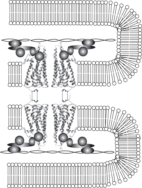
Figure 3.
Hypothetical arrangement of anchoring and tethering mechanisms within rhabdomeric receptors. Visual pigments are arranged in dimers, which are anchored to each other across microvillar membranes (represented by dashed lines), and tethered to cytoskeletal elements (actin core represented by double helix in the middle of the microvilli) by phototransduction molecules (represented by shaded forms).

