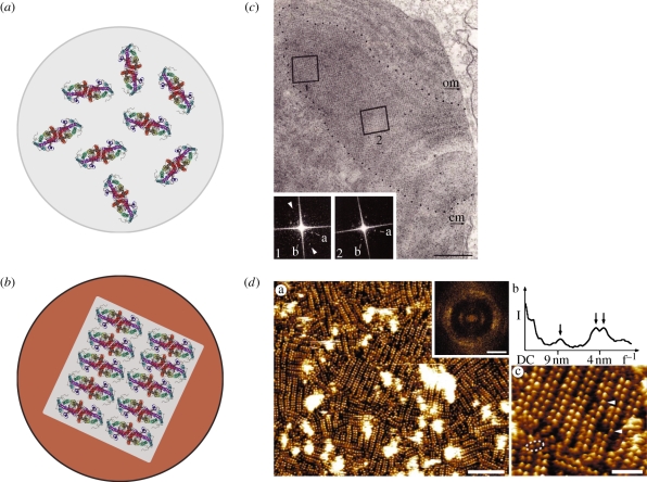
Figure 5.
Potential lipid and protein interactions in ciliary photoreceptors. Schematics of (a) rod discs with opsin dimers still free to rotate versus (b) cone discs with phase separation into cholesterol-rich and more ordered (Lo, blue) and cholesterol-poor and more fluid (Lα, grey) domains, with dimers ordered into rows. Evidence for membrane phase separation from (c) cone transmission electron microscopy studies (adapted from [61]) and (d) rod AFM and x-ray diffraction studies (adapted from [49]).

