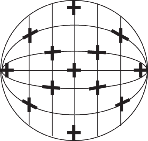
Figure 4.

Representation of the hemisphere model eye cup (viewed from the front). The axes of the orthogonally paired rhabdomeres (represented by black crosses at approximately 30° intervals) are aligned along lines of latitude and longitude that have been rotated 90°, as determined by the hemisphere model.
