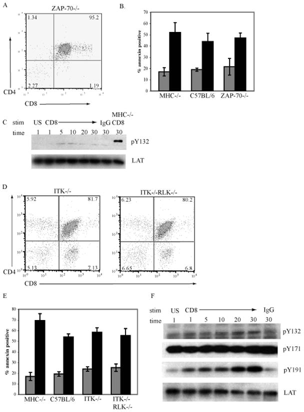
Figure 6. The tyrosine kinase ZAP-70, ITK, and RLK are not required for CD8-mediated apoptosis of DP thymocytes.

A. Thymocytes from ZAP-70−/− mice were stained with antibodies to CD8α and CD4 and analysed by flow cytometry.
B. Thymocytes from MHC−/− C57BL/6, and ZAP-70−/− mice were left unstimulated (grey bars) or treated with antibody to CD8α and crosslinked with goat anti-rat Ig for 6hrs (black bars). The induction of apoptosis was measured by the binding of annexin-V. N=3; mean ± SEM.
C. ZAP-70−/− thymocytes were treated with antibody to CD8α and crosslinked with goat anti-rat Ig for the indicated times, treated only with goat anti-rat Ig or left unstimulated (US). Western blots of whole cell lysates were analyzed for expression of pLATY132 and reprobed with antibodies to LAT as a loading control. MHC−/− cells treated with antibody to CD8α and crosslinked with goat anti-rat Ig served as a positive control.
D. Thymocytes from ITK−/− and ITK−/−RLK−/− mice were stained with antibodies to CD8α and CD4 and analysed by flow cytometry.
E. Thymocytes from MHC−/−, C57BL/6, ITK−/− and ITK−/−RLK−/− mice were left unstimulated (grey bars) or treated with antibody to CD8α and crosslinked with goat anti-rat Ig for 6hrs (black bars). The induction of apoptosis was measured by the binding of annexin-V. N=3; mean ± SEM.
F. ITK−/−RLK−/− thymocytes were treated with antibody to CD8α and crosslinked with goat anti-rat Ig for the indicated times, treated only with goat anti-rat Ig or left unstimulated (US). Western blots of whole cell lysates were analyzed for expression of pLATY132, pLATY171. pLATY191 and reprobed with antibodies to LAT as a loading control.
