Figure 2.
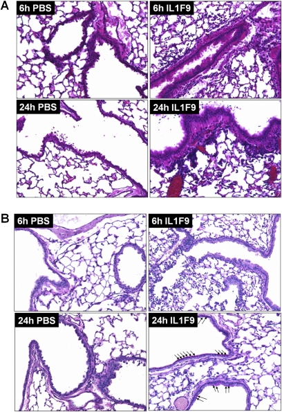
Intratracheal challenge with IL-1F9 leads to cellular infiltrates and mucus production in airways of mice previously unexposed to allergens. (A) Hematoxylin and eosin staining of paraffin-embedded mouse lung sections collected 6 hours and 24 hours after the last of four intratracheal challenges with PBS or IL-1F9. (B) Periodic acid–Schiff staining of paraffin-embedded mouse lung sections collected 6 hours and 24 hours after the last of four intratracheal challenges with PBS or IL-1F9. Arrows indicate PAS positive, mucus-producing cells. Slides are representative of n = 4–5 mice/group/time point.

