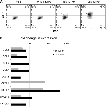
Figure 7.
IL-1F9 stimulated NF-κB activity and induced mRNA expression of neutrophil chemoattractants in mouse macrophage cell lines. (A) RAW264.7 cells stably transfected with NF-κB–responsive E-selectin promoter driving enhanced green fluorescent protein (GFP) expression were stimulated with increasing doses of IL-1F9 for 6 hours. NF-κB activity was measured by the percentage of cells that expressed enhanced GFP after stimulation. A representative depiction is based on two independent experiments with similar results. (B) Murine alveolar macrophage (MH-S) mouse alveolar macrophage cell lines were stimulated with 10 μg IL-1F9 for 1 hour and 6 hours. RNA isolated from cells was used to measure chemokine and chemokine receptor mRNA expression, using a quantitative PCR miniarray. Data were analyzed using the GPR algorithm. Only genes with significant changes in expression (P < 0.05) after IL-1F9 treatment compared with control samples are presented here, as fold change in mRNA expression.

