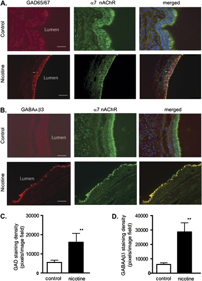
Figure 4.
Chronic nicotine exposure increased GAD and GABAA receptor β3 expression in monkey BECs. (A) Double-labeling with GAD65/67 (red) and α7 nAChR (green) was performed in lung sections of control monkey and nicotine-exposed lungs. Calibration bars represent 45 μM in control lung sections, and 35 μM in nicotine lung sections. (B) Double-labeling with GABAA receptor β3 (red) and α7 nAChR (green) was evident in lung sections of control and nicotine-exposed lung. Arrow indicates a neuroepithelial cell cluster. Calibration bars represent 35 μM. (C and D) Quantitative analysis of immunofluorescence staining, expressed as fluorescent pixels per image field. (C) Immunofluorescence showed an increase in intensity of GAD65/67 in the lung epithelium (control, 5,500 ± 2,000 pixels2; nicotine-exposed lung, 16,013 ± 8,000 pixels2). (D) Immunofluorescence showed an increase in intensity of GABAA receptor β3 in the lung epithelium (control, 6,000 ± 2,000 pixels2; nicotine-exposed lung, 28,630 ± 11,000 pixels2). n = 8 image fields of four lung slices from four monkeys. **P < 0.001.

