Figure 5.
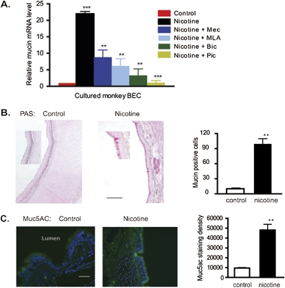
Interactions of nicotine and GABA signaling affect the expression and secretion of mucin. (A) Real-time PCR analysis of mucin 5A expression in cultured BECs treated with nicotine (1 μM for 48 hours) plus the nAChR antagonists mecamylamine (Mec; 25 μM,) and methyllycaconitine (MLA; 30 nM), and the GABAA receptor antagonists, picrotoxin (Pic; 30 μM) and bicuculline (Bic; 30 μM). n = 18 from three experiments. *P < 0.05 for nicotine + antagonist groups compared with nicotine alone group. **P < 0.01 for nicotine-treated group compared with control group. (B) Periodic acid–Schiff (PAS) staining shows increased expression of mucin in nicotine-exposed lung compared with control lung. Quantitation of PAS-positive cells from control and nicotine-exposed animals is shown at right (three animals per group, four sections per animals). **P < 0.01. (C) Immunohistochemistry shows increased expression of mucin-5AC in nicotine-exposed animals. Quantification of immunostained pixels per image field involved three animals per group, and four sections per animal. **P < 0.01.

