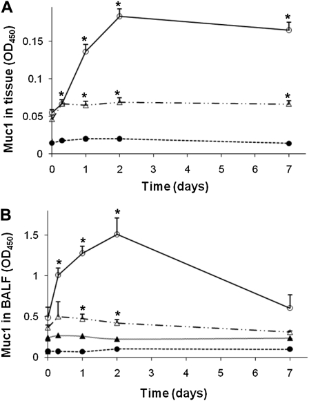
Figure 4.
Muc1 levels of the lung after PA infection. Different strains of mice were infected intranasally with 107 CFU of PAK. At the indicated times, animals were killed and the amounts of Muc1 in the whole lung (A) and the BALF samples (B) were measured by ELISA using anti-MUC1 antibody. CT33 antibody recognizing the cytoplasmic domain of Muc1 was used in (A), whereas F-19 recognizing the extracellular domain of Muc1 was used in (B), as described in Materials and Methods. Open circles, WT; closed circles, Muc1 KO; open triangles, TNFR1 KO; closed triangles, WT by CT33. Notice that Muc1 present in BALF reacts with F-19, but not CT33, indicating that it lacks the CT domain. Each data point represents a mean (± SEM of four samples obtained from four different mice. The results are representative of two separate experiments. *P < 0.05.

