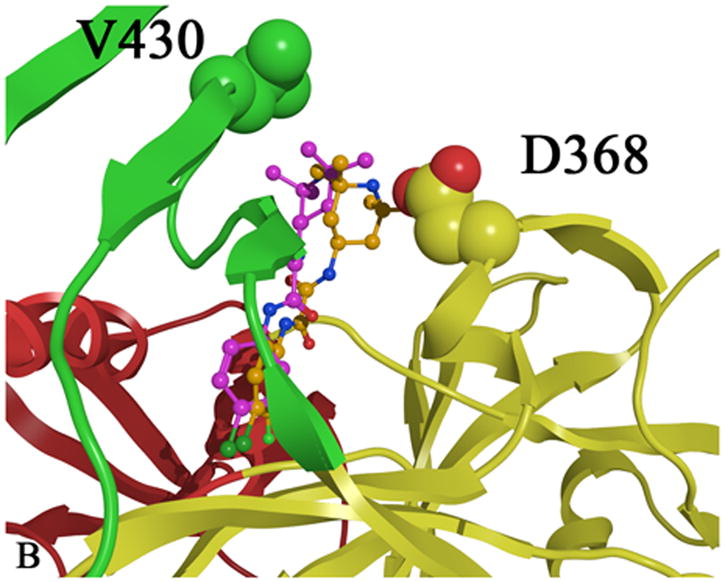
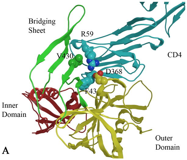
Figure 1.

A) CD4 bound (cyan) to gp120 with the gp120 domains colored as follows: inner domain (red); outer domain (yellow) and bridging sheet (green). Carbon atoms are colored by domain color while non-carbon atoms are colored as follows: oxygen (red) and nitrogen (blue). B) Two plausible docked conformations of 1 (orange and purple) bound in the Phe 43 cavity of gp120 (1G9M). Docking indicates that the p-chloro-m-fluoro-benzenyl binds at the bottom of the Phe 43 cavity, while the tetramethyl-piperidine forms hydrophobic interactions in the vestibule of the cavity between D368 and V430.

