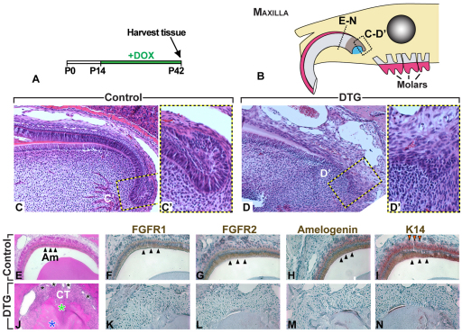
Fig. 4.
Long-term postnatal reduction of FGFR2b signaling leads to defective amelogenesis in maxillary incisors. (A) Timecourse for doxycycline treatment. (B) Dotted lines in schematic indicate areas shown in sagittal sections in C-D′ and planes of coronal sections in E-N. Blue represents mesenchyme and dark brown represents epithelium. (C-D′,E,J) Hematoxylin and Eosin staining of maxillary incisor sections from control (C,C′,E) and DTG (D,D′,J) mice. C′ and D′ are magnifications of the areas indicated in C and D, respectively. Note defects in the dentin and enamel matrices (blue and green asterisks, respectively), ectopic blood vessels (small black asterisks), and connective tissue (CT) in the proximal incisor region (J) where ameloblasts are normally found in controls (E). (F-I,K-N) Immunostaining of coronal sections with antibodies against FGFR1 (F,K), FGFR2 (G,L), amelogenin (H,M) and K14 (L,N). Black arrowheads point to ameloblasts.

