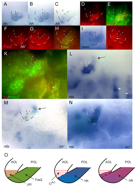
Fig. 4.
Hh signaling in Drosophila embryonic visual primordia. (A-N) Lips of the optic lobe (OL) placode are outlined with dotted lines. Anterior is to the left, dorsal is at the top. Patterns of hh expression in the OL change in early (A), mid (B) and late (C) stage 11 embryos. Fas2 expression (D; red) marks the posterior optic lobe (POL) and is coincident with hh-lacZ at mid-stage (E; green). Fas2 staining (F; red) marks POL in hhAC (hh–). Full-length Ci (G; red) in the AOL and in Fas2-containing POL (H; green). (I) smo expression in AOL and POL. (J) Ptc protein (arrow) in AOL. (K) En (red) is absent from the OL but is present elsewhere where hh is expressed (arrow). (L) rdx expression in POL, in adjacent AOL and in cells dorsal to the OL (black arrow) and in ectodermal stripes (white arrows). (M) rdx expression in hhAC (hh–) is largely absent from the OL but is present in more dorsal cells (arrow). (N) rdx expression in enl11/en7 is absent from ectodermal stripes but unaffected in the OL. (O) Diagram of Hh target gene activation in the optic primordium. Left: Fas2 (green) in the POL and Ptc protein in the adjacent AOL (brown). Middle: CiAct (red) in both the POL and adjacent AOL and Hh (blue) in the POL. Right: rdx (purple) in the POL and adjacent AOL.

