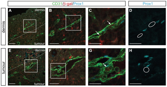
Fig. 3.
Myeloid cells do not comprise a pool of lymphatic endothelial progenitors during tumour-stimulated lymphangiogenesis. Lineage tracing in adult LysMCre+/–;ROSA26R+/– mice revealed select β-galactosidase-positive cells derived from the myeloid lineage apparently integrated within PROX1-positive, CD31-positive pre-existing dermal (A-D) or newly generated peri- or intra-tumoural (E-H) lymphatic vessels. PROX1 expression was not observed in any β-galactosidase-positive cells within lymphatic vessels (C,G, arrows). White circles in D and H illustrate the location of macrophages (arrows) in C and G, respectively. D and H illustrate single-channel PROX1 images of C and G, respectively. Data are representative of eight independent experiments per tumour type. Illustrated is an example of the LLC tumour. Scale bars: 100 μm in A,E; 50 μm in B,F; 25 μm in C,D,G,H.

