Figure 5.
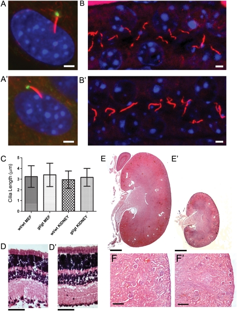
No loss of ciliation is seen Ift80gt/gt animals. (A and A′) Immunofluorescence of MEF cells. Cells are stained with anti gamma tubulin (green) and anti-acetylated alpha-tubulin (red) and nuclei were marked with DAPI (blue). Cilia are present on both wild-type (A) and Ift80gt/gt (A′) cells. Scale bar = 2 µm. (B and B′) Immunofluorescence of P21 kidney sections. Sections are stained with anti-acetylated alpha-tubulin (red) and nuclei were marked with DAPI (blue). Cilia are present in both wild-type (B) and Ift80gt/gt (B′) tubules. Scale bar = 2 µm. (C) Length analysis of both MEF and tubule cilia. Five hundred wild-type and Ift80gt/gt MEF cilia were measured as were 200 wild-type and Ift80gt/gt kidney cilia. The data show no significant difference between the lengths of cilia in either genotype. Error bars represent one standard deviation from the mean. (D and D′) Haematoxylin and eosin staining of wild-type (D) and Ift80gt/gt (D′) retina at P21. All retinal layers are present in the Ift80gt/gt. Scale bar = 100 µm. (E and F′) Haematoxylin and eosin staining of wild-type (E and enlargement F) and Ift80gt/gt (E′ and enlargement F′) kidneys at P21. Ift80gt/gt kidneys are smaller in line with the size of the mouse but appear normally developed with mature glomeruli and no evidence of cysts. Scale bars = 500 µm (E and E′) and 125 µm (F and F′).

