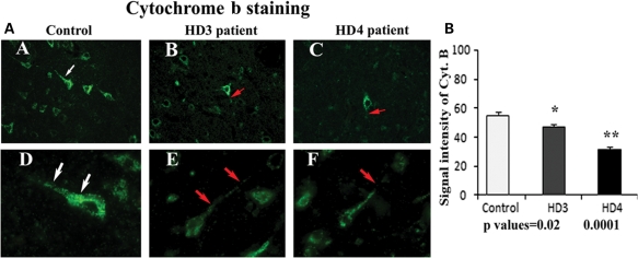
Figure 5.
(A) Mitochondrial-encoded proteins and cytochrome b immunoreactivity in frontal cortex sections from grade III (B) and grade IV HD (C) patients and controls (A). Uniform immunoreactivity of cytochrome b was found in the control subject (D). Interrupted immunoreactivity of cytochrome b (red arrows) was found in grade III (E) and grade IV (F) HD patients. Arrows indicate immunoreactivity. Images were photographed at ×40 (upper panel) and ×100 (lower panel). (B) Quantitative analysis of cytochrome b. Significantly decreased cytochrome b in grade III (P< 0.02) and grade IV (P< 0.0001) HD patients. Error bars indicate mean ± SEM.

