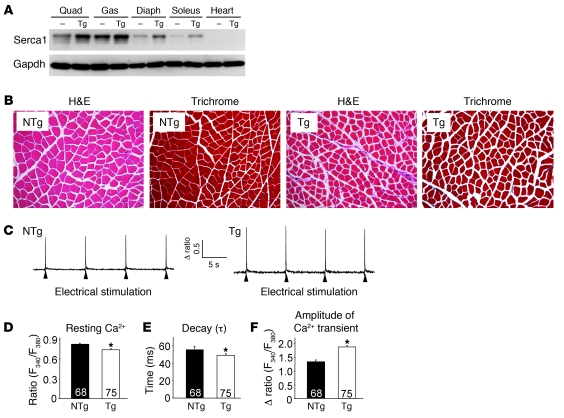
Figure 1. Overexpression of SERCA1 in skeletal muscle enhances Ca2+ cycling during EC coupling.
(A) Western blot analysis for SERCA1 expression in different muscle groups isolated from non-Tg (NTg) and SERCA1 Tg (Tg) mice at 3 months of age. Quad, quadriceps; Gas, gastrocnemius; Diaph, diaphragm. (B) H&E and Masson’s trichrome sections of quadriceps. Original magnification, ×200. (C) Representative traces of F340/F380 fluorescence ratio recordings from single FDB myofibers isolated from NTg and SERCA1 Tg mice in response to electrical stimulation. (D) Resting Ca2+ ratio, (E) time constant of decay (τ), and (F) peak Ca2+ transient amplitude in isolated myofibers from the indicated genotypes. *P < 0.05 compared with NTg mice; n = total number of fibers recorded from 4 animals in each genotype shown in the graphs, D–F.

