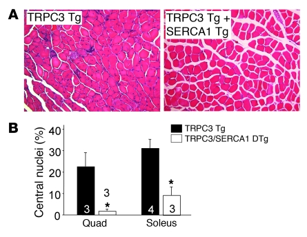
Figure 4. SERCA1 mitigates histological features of MD in TRPC3 Tg mice.
(A) Representative histological H&E stain of quadriceps from TRPC3 Tg and TRPC3/SERCA1 double-Tg at 3 months of age. Original magnification, ×200. (B) Percentage of myofibers with centrally located nuclei in quadriceps and soleus from TRPC3 Tg and TRPC3/SERCA1 double-Tg mice. At least 3 mice from each genotype were used. *P < 0.05 versus TRPC3 TG. Number of mice used is shown in the graph.

