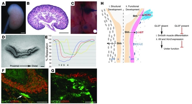
Figure 3. Elimination of GLI3 repressor in the Smo-deficient background rescues the renal phenotype.
(A–G) Analysis of Rarb2-Cre;SmoloxP/–;Gli3XtJ/XtJ embryos at E18.5 revealed normalization of kidney and ureter morphology (A–C), rescue of coordinated proximal-distal ureter contractions (D and E; blue line [1], proximal ureter; red line [2], midproximal ureter; green line [3], mid-distal ureter; yellow line [4], distal ureter), and restoration of Kit (F) and Hcn3 (G) expression. In E, percent relaxation is plotted for the duration of a single peristaltic movement (x axis). Scale bars: 100 μm (D, F, and G), 500 μm (A–C). (H) Model for a bifunctional role for Hh signaling during the functional development of the ureter. (i) Shh secreted by the ureteric bud/urothelium is required during early development for smooth muscle cell development and the patterning of the ureter mesenchyme. (ii) During later development, SHH-SMO dependent signaling is required for Kit and Hcn3 expression in distinct cell populations that regulate coordinated ureter contractions. SHH signaling to adjacent cells promotes pathway activation and prevents formation of GLI3 repressor (GLI3R). In the presence of GLI3 repressor function during ureter morphogenesis, smooth muscle cell differentiation and expression of Kit and Hcn3 is impaired, leading to abnormal ureter function. u, ureteric bud/urothelium; um, ureter mesenchyme; s, subepithelial layer; sm, smooth muscle; c, outer connective tissue.

