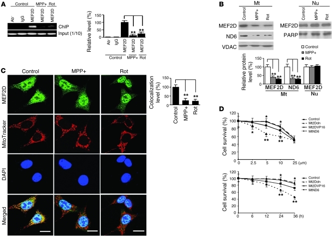
Figure 6. Inhibition of mitochondrial MEF2D by toxic signals relevant to PD.
(A) Reduced binding of MEF2D to ND6 after neurotoxin treatment. SN4741 cells were treated with MPP+ (25 μM) or rotenone (Rot, 100 nM) for 12 hours. ChIP assay showed that binding of MEF2D to ND6 was greatly reduced (n = 4; **P < 0.01). Control indicates untreated. (B) Reduced mitochondrial MEF2D and ND6 protein levels after neurotoxin treatment. Western blotting showed that levels of MEF2D and ND6 in purified mitochondria, but not in nuclei, were significantly reduced (n = 4; **P < 0.01). Control indicates untreated. (C) Immunocytochemical analysis of mitochondrial MEF2D after MPP+ and rotenone treatment. MPP+ and rotenone preferentially reduced colocalization of MEF2D with MitoTracker (n = 50 cells; **P < 0.01). Scale bars: 10 μm. Experiments were repeated 4 times. Control indicates untreated. (D) Effect of mitochondrial MEF2D-ND6 pathway on MPP+ toxicity in SN4741 cells. SN4741 cells were treated with MPP+ after infection with the control or with Mt2Ddn, Mt2DVP16, or MtND6 lentiviruses. Treatment was either with different doses for 24 hours (top) or the 5-μM dose for different times (bottom). Cell viability was measured by WST-1 assay (n = 4; *P < 0.05; **P < 0.01). Control indicates the control vector group.

