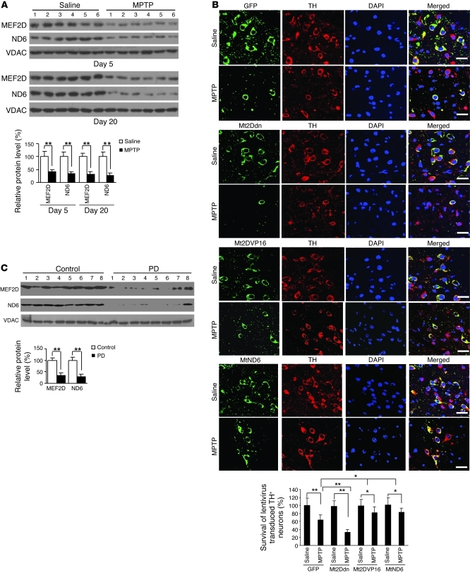
Figure 7. Correlation of mitochondrial MEF2D in a MPTP model of PD and in postmortem brains of PD patients.
(A) Reduced mitochondrial MEF2D and ND6 levels in the brains of MPTP-treated mice (n = 18; **P < 0.01). Mitochondria purified from brain SNpc region were analyzed by Western blotting. Experiments were repeated 3 times. (B) Role of mitochondrial MEF2D-ND6 pathway in maintaining TH+ neurons in SNpc in a MPTP mouse model of PD. For each group, 3 mice received stereotactic injection of control vector (GFP) or Mt2Ddn lentivirus in SN. 2 weeks later, mice were exposed to MPTP. After treatment for 7 days, survival of lentivirus-transduced TH+ neurons in SN was determined by immunohistochemistry. Scale bars: 30 μm. Quantitative analysis of 9 mice from 3 independent experiments is also shown (**P < 0.01). (C) Reduced mitochondrial MEF2D and ND6 levels in the brains of human PD patients. Mitochondria were purified from brain striata of postmortem PD patients and normal controls. Equal amounts of mitochondrial proteins were subjected to Western blotting. Quantitative analysis of the bands is also shown (n = 13 patients and 13 controls; *P < 0.01). Experiments were repeated 2 times.

