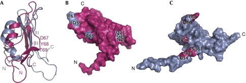
Figure 2.
Structural comparison. (A) Ribbon diagrams of the DaliLite (Holm & Park, 2000) fit of Escherichia coli BamE (grey) to Xanthomonas axonopodis pv. citri BamE (pink; Protein Data Bank number 2pxg; Vanini et al, 2008). An alignment of this fit can be seen in supplementary Fig S2 online. Residues 67–69 of X. axonopodis pv. citri BamE are labelled and form a β1-strand kink not present in the E. coli protein. (B) Surface representation of X. axonopodis pv. citri BamE showing the positions of the QGN motif and the highly conserved Pro 55, Asp 59 and Phe 61 (shown in grey). (C) Similar view of E. coli BamE showing the positions of the homologous residues in pink and highlighting the distinct position of the QGN motif. BamE, β-barrel assembly machine E.

