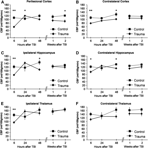
Figure 3.
Regional cerebral blood flow (CBF) quantified by arterial spin labeling (ASL) magnetic resonance imaging (MRI) over 14 days after traumatic brain injury (TBI). (A) Perilesional cortex, (B) contralateral cortex, (C) ipsilateral hippocampus, (D) contralateral hippocampus, (E) ipsilateral thalamus, and (F) contralateral thalamus. Data are shown as mean±s.e.m. The data at 6 hours come from 6 controls and 15 rats with TBI. For the analysis at later time points, there were four controls and nine TBI rats. Statistical significances: *P<0.05, **P<0.01 (Mann–Whitney U-test, control versus TBI at the same time point). Comparison of ipsilateral changes with those contralaterally (Wilcoxon's test) indicated that at 6 hours after injury, ipsilateral CBF was lower than contralateral CBF in the perilesional cortex, hippocampus, and thalamus (P<0.01). Twenty-four hours after injury, ipsilateral CBF was elevated compared with contralateral CBF in the perilesional cortex (P<0.01), hippocampus (P<0.05), and thalamus (P<0.01). Two days after injury, ipsilateral CBF was lower than contralateral CBF in the hippocampus (P<0.05) and thalamus (P<0.01). Compared with the contralateral cortex, CBF in the perilesional cortex became lower 1 week after injury (P<0.05) and remained so thereafter. At 1 and 2 weeks after injury, thalamic CBF was slightly greater ipsilaterally than contralaterally (P<0.05). The CBF in controls did not change between time points (Wilcoxon's test).

