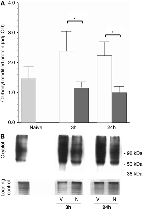
Figure 3.
Protein oxidation is suppressed by necrostatin treatment. (A) Carbonyl-modified protein in the forebrain from naive animals (light gray) and at 3 and 24 hours after hypoxia–ischemia (HI) and treatment with vehicle (white) or necrostatin (dark gray). Data represented as bars (mean±s.e.m.). *P<0.05. (B) Representative OxyBlots with corresponding loading controls are shown for vehicle (V) and necrostatin (N) at 3 and 24 hours after HI and treatment. Position of molecular weight standards is shown for reference.

