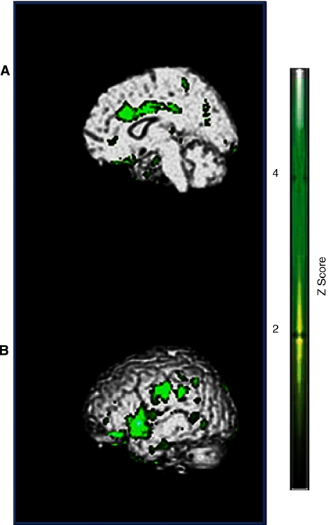
Figure 1.
(A) Limbic lobe, anterior cingulum. (B) Superior temporal gyrus, parietal lobe, postcentral gyrus. Hypoperfused brain regions in PET scan study: green areas correspond to hypoperfusion in patients with PWS versus controls (P<0.05 uncorrected). PET, positron emission tomography; PWS, Prader–Willi syndrome.

