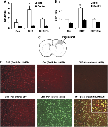
Figure 2.
Salt-induced kinase 1 (SIK1) expression was induced in rat brains at 12 hours reperfusion after middle cerebral artery occlusion (MCAO). The SIK1 mRNA was significantly increased in ipsilateral (Ipsil) cortical penumbra (A) and striatal penumbra (B) of dihydrotestosterone (DHT) (15 mg)-supplemented castrates (n=5) compared with unsupplemented castrates (Cas, n=6). In DHT plus flutamide-treated castrates (DHT+Flu, n=3), DHT induction of SIK1 mRNA was attenuated by flutamide. *P<0.05 versus Cas and DHT+Flu; Contra: contralateral side. (C) Schematic drawing of a rat coronal brain slice illustrating the region (shaded area) for immunohistochemistry analysis. (D) Immunohistochemistry analysis of SIK1 induction. At 12 hours reperfusion, SIK1 protein (red) was induced in cortical peri-infarct regions of DHT-supplemented rats compared with contralateral sides; and SIK1 protein was translated in the peri-infarct of castrates (upper panel). The SIK1 immunofluorescence predominantly colocalized with the neuronal marker NeuN (green, lower panel). The inset in the lower right image showed SIK1 (red) and NeuN (green) colocalization at a higher magnification. The color reproduction of this figure is available on the html full text version of the article.

