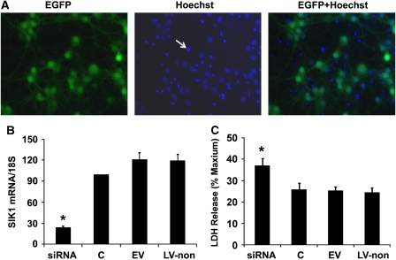
Figure 3.
Salt-induced kinase 1 (SIK1) knockdown exacerbated oxygen–glucose deprivation (OGD)-induced neuronal death. (A) Photomicrographs of primary neurons infected with lentivirus. At 4 days after infection, neurons were positive for enhanced green fluorescence protein (EGFP) (the left image: green fluorescence). Lentivirus-infected neurons were further stained with a nuclear dye Hoechst 33258 (the center image: blue fluorescence); and eGFP fluorescence, which indicated lentiviral infection, almost completely colocalized with Hoechst staining of live cells (the right image). In the center and right images, densely packed Hoechst staining, as pointed by an arrow in the center image, indicated the nuclei of dead cells. Cell death was not induced by lentiviral infection as control neurons without lentiviral infection at day 10 in vitro also displayed similar percentages of densely packed Hoechst staining. (B) Lentivirus-expressing SIK1 small interference RNA (siRNA) decreased SIK1 mRNA by 75% in primary neurons compared with control neurons without lentiviral infection or neurons infected with EV or LV-non (C, control neurons without lentiviral infection; EV, empty virus-expressing eGFP only; LV-non, lentivirus-expressing nonsense siRNA). (C) The SIK1 knockdown by lentivirus-delivered siRNA significantly increased cell death induced by 2.5 hours OGD in primary cortical neurons. Cell death was measured with lactate dehydrogenase (LDH) release assay at 24 hours reoxygenation. Results are expressed as ratios to maximum release when all cells are killed by triton. eGFP. *P<0.05 versus C, EV, or LV-non (n=7). The color reproduction of this figure is available on the html full text version of the article.

