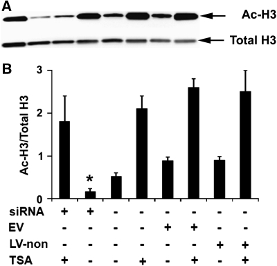
Figure 5.
Salt-induced kinase 1 (SIK1) knockdown decreased basal histone H3 acetylation but did not decrease trichostatin A (TSA)-elevated basal H3 acetylation levels in primary neurons. Representative images of Western blot analysis of basal acetylated (Ac-H3) and total histone H3 (Total H3) in primary neurons at the time point when cultures were exposed to oxygen–glucose deprivation (OGD) insults (A) and bar graph of the Western blot results (B). C, control neurons without lentiviral infection; EV, neurons transduced with empty virus-expressing enhanced green fluorescence protein (eGFP) only; LV-non, neurons infected with lentivirus-expressing nonsense small interference RNA (siRNA) and eGFP. *P<0.05 versus C, EV, or LV-non in the absence of TSA (n=5).

