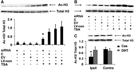
Figure 7.
Salt-induced kinase 1 (SIK1) knockdown decreased trichostatin A (TSA)-elevated histone H3 acetylation after oxygen–glucose deprivation (OGD) and dihydrotestosterone (DHT) prevented ischemia-induced loss of histone H3 acetylation after middle cerebral artery occlusion (MCAO). (A) Representative image of Western blot analysis of acetylated (Ac-H3) and total histone H3 (Total H3) in primary neurons at 4 hours reoxygenation after 2.5 hours OGD and bar graph of the Western blot results. *P<0.05 versus C, EV, or LV-non in the presence of TSA (n=3; C, control neurons without lentiviral infection; EV, empty virus-expressing enhanced green fluorescence protein (eGFP) only; LV-non, lentivirus-expressing nonsense small interference RNA (siRNA) and eGFP). (B) Image of Western blot analysis of histone H3 acetylation patterns in normoxic control neurons, which were not different from basal histone H3 acetylation patterns (as shown in Figure 5). (C) Representative image of Western blot analysis of acetylated and total histone H3 in the rat brain at 12 hours reperfusion after MCAO and bar graph of the Western blot results. Compared with castration, DHT at the protective dose prevented ischemia-induced loss of histone H3 acetylation in ipsilateral cortical peri-infarct regions. *P<0.05 versus Cas (Ipsil), DHT (Contra), and Cas (Contra) (n=3).

