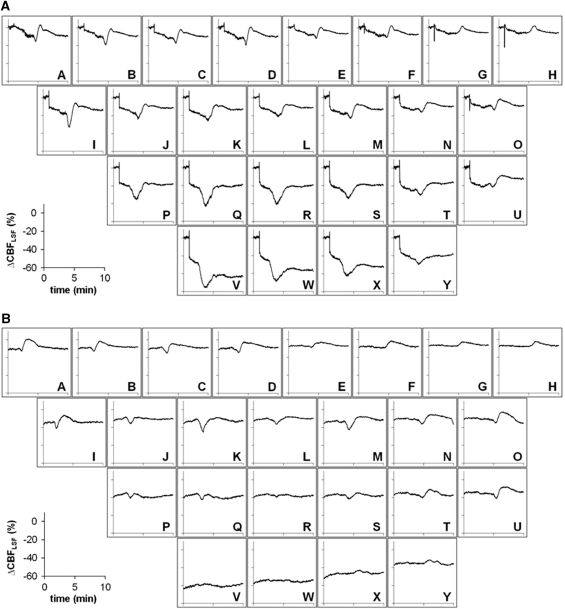
Figure 5.
Examples of line plots of a primary concentric and a secondary circumferential CBFLSF wave analyzed over 10 minutes for the whole set of 25 individual regions of interest (ROIs). (A) The primary concentric wave appeared in each ROI showing monophasic hypoemic wave morphology in frontolateral ROIs (VWX and PQR) and biphasic or monophasic hyperemic morphology in medial ROIs. (B) The secondary circumferential wave showed a similar wave morphology with respect to the CBF gradient within the ischemic focus, but CBFLSF waves were not observed in the core of the ischemic territory.

