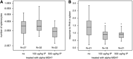
Figure 4.
Treatment with α-MSH decreases splenocyte responsiveness to PHA. (A) There were no significant differences in the numbers of splenocytes at 24 hours after stroke among the different treatment groups. (B) Animals treated with either dose of α-MSH, however, had decreased responsiveness of the splenocytes to PHA at this time point. Box plots show median, IQR, and outliers. Differs from control group at *P<0.05 by Mann–Whitney U-test. α-MSH, α-melanocyte-stimulating hormone; IQR, interquartile range.

