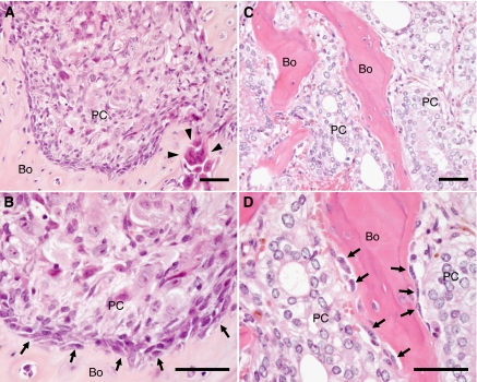
Figure 1.
Histological findings suggesting direct prostate cancer-cell-osteoblast contact in bone metastases. The indicated tissues were fixed and analysed following H&E staining. (A and B) An osteolytic lesion produced by the growth of PC-3 human prostate cancer cells in the tibia of a nude mouse. The close proximity of PC-3 cells to osteoblasts suggesting direct cancer-cell-osteoblast contact, as well as osteolysis by osteoclasts was observed. (C and D) Representative osteoblastic bone metastases of autopsy cases of prostate cancer. The close proximity of prostate cancer cells to osteoblasts suggesting direct cancer-cell-osteoblast contact was also observed. Arrows, osteoblasts; arrow heads, osteoclasts; Bo, bone; PC, prostate cancer cells. Scale =50 μm.

