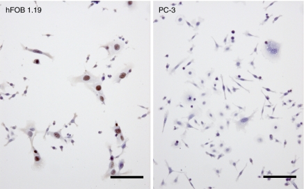
Figure 2.
Immunohistochemical analysis of the SV40 large T antigen after sorting of cells in contact cocultures. PC-3 human prostate cancer cells (labelled with DiOC18(3)) were mixed and cocultured with DiIC18(3)-labeled human osteoblasts expressing SV40 (hFOB1.19) for 48 h. Cocultured prostate cancer cells (DiOC18(3)+/DiIC18(3)−) and hFOB1.19 cells (DiOC18(3)−/DiIC18(3)+) were then separated using flow cytometry. One thousand cells from each sorted cell population were cultured overnight on glass slides and were then immunohistochemically stained for the SV40 large T antigen. Positive staining of the SV40 large T antigen was observed in the nuclei of hFOB1.19 cells (DiOC18(3)−/DiIC18(3)+) but not in PC-3 cells (DiOC18(3)+/DiIC18(3)−). Scale =100 μm.

