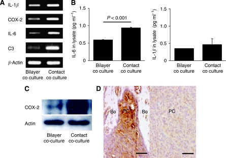
Figure 3.
Analysis of IL-1β, COX-2, IL-6 and C3 expression in PC3 cells cocultured with hFOB1.19 cells. (A) The mRNA expression of the four osteoclastogenesis-related genes, IL-1β, COX-2, IL-6 and C3, in PC-3 cells following coculture under contact and bilayer conditions, was analysed using RT–PCR. Consistent with the cDNA microarray results, these genes were upregulated in contact cocultured PC-3 cells compared with bilayer cocultured cells. The primers used for RT–PCR are shown in Table 1. (B) Measurement of IL-6 and IL-1β levels in PC-3 cells using ELISA. The level of IL-6 was significantly higher (P<0.001) in PC-3 cells under contact coculture conditions than under bilayer coculture conditions, whereas IL-1β levels did not significantly differ between the two coculture conditions. ELISA was performed using a PC-3 cell lysate. (C) Analysis of COX-2 expression in PC-3 cells by western blotting. Expression of COX-2 was higher in PC-3 cells under contact coculture conditions than under bilayer coculture conditions. (D) Immunohistochemical analysis of COX-2 expression in an osteolytic bone metastasis model. Left: Strong expression of COX-2 in PC-3 cells adjacent to the bone. The close proximity of PC-3 cells to osteoblasts suggesting direct cancer-cell-osteoblast contact was observed in this area. Right: very low expression of COX-2 in PC-3 cells distant from the bone. Bo, bone; PC, prostate cancer cells. Scale=50 μm.

