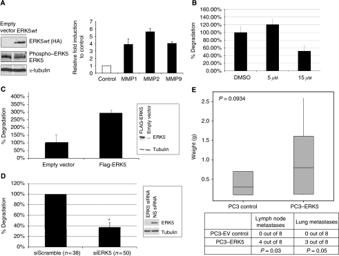
Figure 3.
(A) Extracellular signal-regulated protein kinase 5-induced MMP promoter activity – (left panel) ERK5 DNA was transiently transfected into PC3 cells where a band (*) corresponding to phospo-ERK5 was seen on stimulation with EGF. (right panel) PC3 cells were co-transfected with MMP1, MMP2 or MMP9 promoter luciferase constructs along with ERK5 or empty plasmid. Transfection efficiency was assessed and normalised to β-galactosidase activity. Data represent mean fold induction compared with basal level of promoter activity from sets of quadruplicate assays±s.e. Experiments were performed three times. (B) Invadopodia formation by A375MM cells is significant suppressed by treatment with 15 μM (but not 5 μM) PD184352 (P<0.005). (C) Transfected ERK5 significantly drives invadopodia formation in PC3 cells (P<0.005). Insert of western blot shows upregulated ERK5 expression in PC3 cells. (D) Invadopodia formation in A375MM cells is significantly suppressed by siRNA-mediated knockdown of ERK5 expression (*P<0.05). Control cells were transfected with non-silencing scrambled siRNA. Number in brackets denotes the cells studied for each condition. Insert of western blot shows reduced ERK5 expression following siRNA transfection. (E) PC3–ERK5 cells form significantly more metastatic lesions (lymph nodes and lung) than control cells in an orthotopic prostate model (P=0.03 and P=0.05 respectively).

