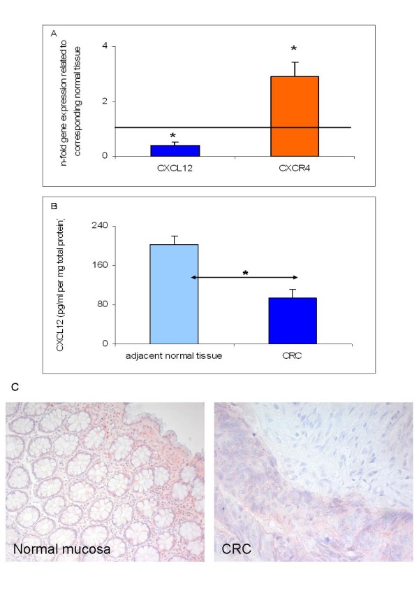
Figure 1.
CXC12/CXCR4 expression in colorectal cancer (CRC) tissue specimens as determined by (A) Q-RT-PCR for CXCL12 and CXCR4, (B) ELISA analysis for CXCL12 and (C) immunohistochemistry for CXCR4. (A) Q-RT-PCR data are expressed as mean +/- standard error of the mean (SEM), *P < 0.05, n = 50. Fold increase above 1 indicates gene overexpression in affected tissues related to unaffected neighbor tissues. (B) Detection of CXCL12 protein concentrations (pg/ml pro mg total protein) in total cell lysates of CRC and adjacent normal tissues from CRC patients (n = 50). Protein data are expressed as mean +/- SEM, *P < 0.05. (C) Detection of CXCR4 protein expression in representative CRC specimens as assessed by immunohistochemical staining with CXCR4-specific antibodies showing positive cytoplasmic staining in CRC and in unaffected corresponding tissues (original magnification × 200 and × 400).

