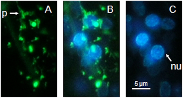
Figure 5. Simultaneous uptake of Alexa Fluor 488-labelled nAChRbp and bisbenzimide by a J2 of Heterodera schachtii.
J2 of H. schachtii were incubated for 16 h in 183 µM nAChRbp labelled with Alexa Fluor 488 together with 1 mM bisbenzimide. The fluorophores were visualised by different excitation and emission wavelengths. Images correspond to a region 5–30 µm posterior to the nerve ring. A) Green fluorescence of the labelled peptide. C) Blue fluorescence of bisbenzimide. B) Combined image of A) and C). Key: p, process of a neuron; nu, nucleus. The scale bar is 5 µm and the images are left lateral views with the anterior uppermost.

