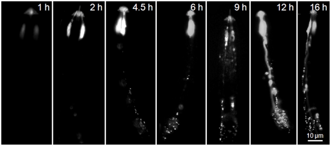
Figure 6. Neuronal uptake of Alexa Fluor 488-labelled nAChRbp by J2s of Globodera pallida.
J2 of G. pallida were incubated in 281 µM nAChRbp labelled with Alexa Fluor 488 for varying periods of 1–16 h and the labelled peptide was then visualised by epifluorescence. Images with one amphidial pouch visualised are lateral views and those with two amphidial pouches visualised are dorsal or ventral views. The scale bar of 10 µm applies to all images.

