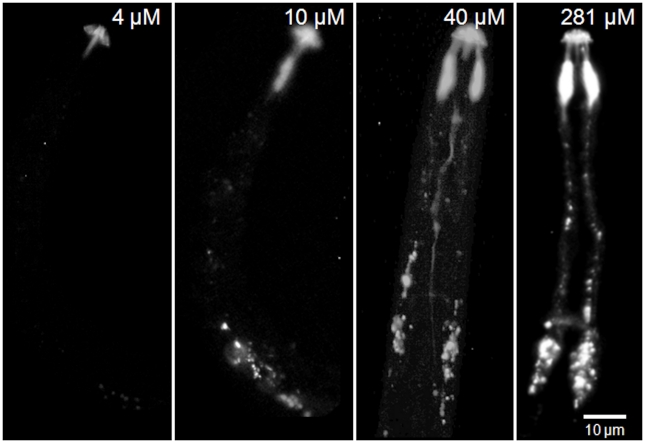
Figure 7. Neuronal uptake of four different concentrations of labelled nAChRbp by J2s of Globodera pallida.
J2 of G. pallida were incubated for 16 h in 4 µM, 10 µM, 40 µM or 281 µM nAChRbp labelled with Alexa Fluor 488. The peptide was visualised by epifluorescence. The scale bar of 10 µm applies to all images.

