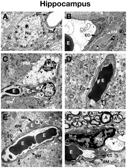
Figure 2. Electron microscope examination of the hippocampus of Naglu mutant mice.
(A), (B) Hippocampal area of C57 BL/6J control mouse shows typical normal ultrastructure of neurons and all capillary components. (C) Vacuolated endothelial cells and pericytes can be observed in 3 months old Naglu mouse. A highly vacuolated perivascular macrophage was also noted. Platelets attracted to a region of endothelial cell damage are visible. (D) Perivascular space can be observed around the capillaries in the hippocampus of an early symptomatic mouse at 3 months of age. (E) In Naglu mouse at 6 months of age, a microaneurysm is visible in the hippocampus, adjacent to a ruptured endothelium. In the microaneurysm, an erythrocyte can be seen trapped under the basement membrane, portending imminent aneurysm rupture. (F) Under high magnification, distended nucleus of pericyte with large vacuoles can be observed in late symptomatic mutant mouse. EC - endothelial cell, BM - basement membrane, E – erythrocyte, m – mitochondrion, A – axon, N – neuron, P – pericyte, PM – perivascular macrophage, Nu – nucleus, V – vacuole, Ma – microaneurysm, pl – platelets, asterisks - microvilli, > - extracellular edematous space. Magnifications: (A), (B), (D), (E): 7,100x; (C): 3,500x; (F): 8,900x.

