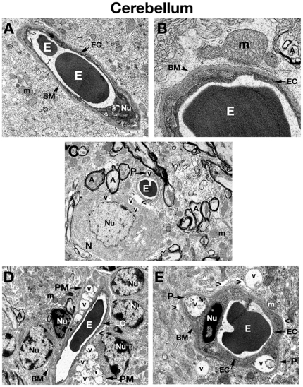
Figure 4. Electron microscope examination of the cerebellum of Naglu mutant mice.
(A), (B) Similar to ultrastructure of microvessels in the cerebral cortex, hippocampus, and striatum, the intact BBB in the cerebellum of a C57 BL/6J control mouse consists of an endothelial cell and a single layer of basement membrane. (C) Edematous space around vessel as well as vacuolated pericyte is found in the cerebellum of 3 months old mutant mouse. Vacuoles are also seen in the cytoplasm of neuron located near vessel. (D) In 6 months old Naglu mice, two highly vacuolated perivascular macrophages surround a microvessel. Edematous space around vessels is in contact with astrocytic end-feet. (E) Pericytes, which contain large vacuoles displacing and destroying cell organelles, can be seen under the capillary basement membrane in mouse at the same stage of disease. Large vacuoles are also visible outside vessel. EC - endothelial cell, BM - basement membrane, E – erythrocyte, m – mitochondrion, A – axon, N – neuron, P – pericyte, PM – perivascular macrophage, Nu – nucleus, V – vacuole, > - extracellular edematous space. Magnifications: (A) 7,100x; (C), (D): 4,400x; (B): 28,000x; (E): 11,000x.

