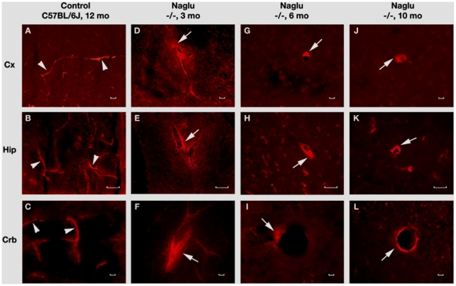
Figure 5. Evans Blue (EB) fluorescence in the brains of Naglu mice.
In the brain, EB can be clearly detected within the blood vessels (A, B, C, red, arrowheads) in the control C57 BL/6J mouse at 12 months of age. In Naglu mice, vascular leakage of EB (red, arrows) is visible in various brain structures (D, E, F) at early (3 months of age), (J, H, I) at late (6 months of age), and (J, K, L) at end stage of disease (10 months of age). Significant EB diffusion into the brain parenchyma from many blood vessels can be detected in end-stage Naglu mouse (10–12 months of age). Cx – cerebral cortex, Hip – hippocampus, Crb – cerebellum. Scale bar in A through L is 25 µm.

