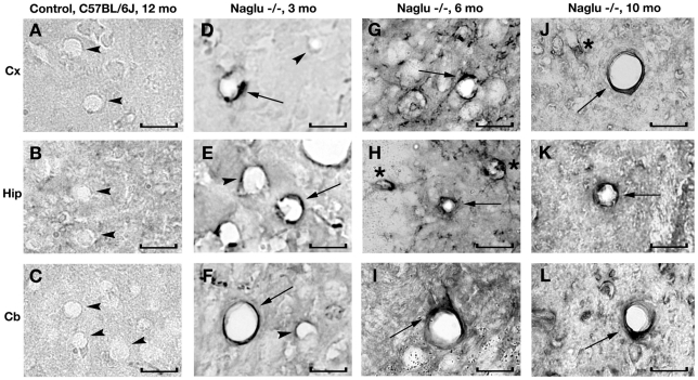
Figure 7. Immunohistochemical staining for GM3 ganglioside in the brains of Naglu mice.
In the brain of control C57 BL/6J mouse at 12 months of age, GM3 ganglioside is not detected within the vascular endothelium of blood vessels (A, B, C, arrowheads). In early symptomatic (3 month of age) Naglu mice, some endothelial cells stained positive for GM3 (D, E, F, arrows) and some cells were negative for GM3 (D, E, F, arrowheads). GM3 accumulation (arrows) was seen in the endothelia of numerous brain blood vessels (G, H, I) at late (6 months of age) and (J, K, L) at end stages of disease (10 months of age). Significant GM3 ganglioside accumulation was also seen in neurons (asterisks) in Naglu mice. Cx – cerebral cortex, Hip – hippocampus, Crb – cerebellum. Scale bar in A through I is 25 µm.

